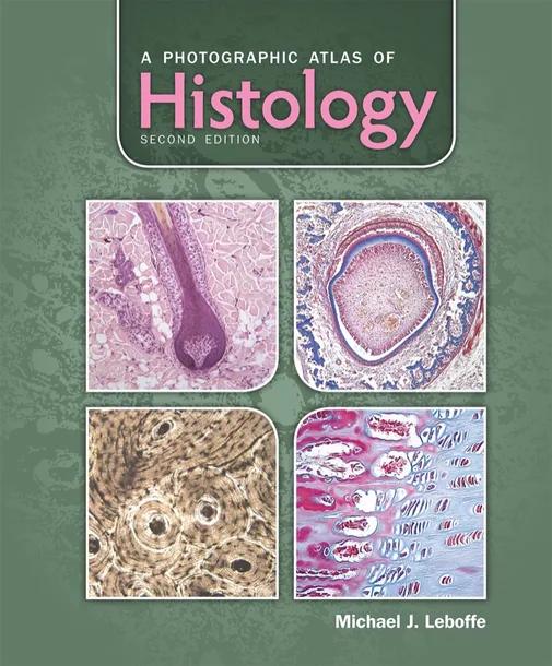
A Photographic Atlas of Histology
by Michael J. Leboffe
2nd Edition
Publisher: Morton Publishing Company
Book Details
| Print ISBN | 9781617310683 |
| eText ISBN | 9781617310683 |
| Publisher | Morton Publishing Company |
| Publishing Year | 2013 |
| Edition | 2nd Edition |
| Language | English |
| Pages | 256 |
A Photographic Atlas of Histology, 2nd Edition, serves as a visual reference intended for undergraduate courses in histology and human anatomy. The publication provides detailed views of human tissues and organs to support academic study and laboratory investigations.
Coverage focuses on primary tissue types and major organ systems across the human body. The atlas examines epithelial, connective, muscular, and nervous tissues, while also addressing complex functional systems such as the cardiovascular, lymphatic, digestive, and respiratory networks.
To assist practical study, the atlas incorporates more than 550 photomicrographs alongside explanatory diagrams and illustrations that clarify histological concepts. The photographic specimens are prepared from slides obtained through biological supply vendors, allowing students to match atlas imagery with what they encounter during laboratory sessions.
Table of Contents
Chapter 1: Introduction
Chapter 2: The Cell
Chapter 3: Epithelial Tissue
Chapter 4: Fibrous Connective Tissues
Chapter 5: Cartilage and Bone
Chapter 6: Blood and Bone Marrow
Chapter 7: Muscle Tissue
Chapter 8: Nervous Tissue and Organs of the Nervous System
Chapter 9: Special Sensory Organs
Chapter 10: Endocrine System
Chapter 11: Integumentary System
Chapter 12: Cardiovascular System
Chapter 13: Lymphatic System
Chapter 14: Digestive System
Chapter 15: Respiratory System
Chapter 16: Urinary System
Chapter 17: Reproductive Systems
Customer Reviews
0.0
0 reviews
No reviews yet. Be the first to review this book!
Write a Review
Reviewed by GradeFocus Editorial Team