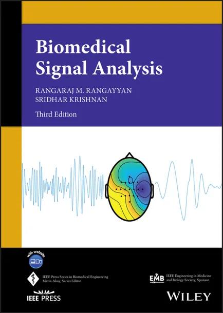
Biomedical Signal Analysis
by Rangaraj M. Rangayyan, Sridhar Krishnan
3rd Edition
Publisher: Wiley-IEEE Press
Book Details
| Print ISBN | 9781119825852 |
| eText ISBN | 9781119825876 |
| Publisher | Wiley-IEEE Press |
| Publishing Year | 2024 |
| Edition | 3rd Edition |
| Language | English |
| Pages | 720 |
Biomedical Signal Analysis, 3rd Edition, is a comprehensive textbook that introduces senior undergraduate and graduate engineering students to digital signal processing methods for computer-aided diagnosis. Early modules covering basic signals, systems, and mathematical transforms establish foundational principles for learners from non-electrical engineering disciplines.
The volume structures its analytical topics around primary clinical objectives. Chapters detail filtering techniques designed to eliminate unwanted artifacts, background noise, and physiological interference, while presenting event detection procedures for locating QRS complexes, heart sounds, and dicrotic notches. Additional sections present the evaluation of concurrent, coupled, and correlated processes, demonstrating these concepts through sleep apnea monitoring applications.
Each chapter opens with a distinct biomedical signal problem statement, followed by real-world case studies and signal illustrations. Learning tools include end-of-chapter study questions, laboratory exercises, and access to an online repository containing problem solutions and empirical data files.
Table of Contents
Chapter 1: Introduction to Biomedical Signals
- • 1.1 The Nature of Biomedical Signals
- • 1.2 Examples of Biomedical Signals
- • 1.2.1 The action potential of a cardiac myocyte
- • 1.2.2 The action potential of a neuron
- • 1.2.3 The electroneurogram (ENG)
- • 1.2.4 The electromyogram (EMG)
- • 1.2.5 The electrocardiogram (ECG)
- • 1.2.6 The electroencephalogram (EEG)
- • 1.2.7 Event-related potentials (ERPs)
- • 1.2.8 The electrogastrogram (EGG)
- • 1.2.9 The phonocardiogram (PCG)
- • 1.2.10 The carotid pulse
- • 1.2.11 The photoplethysmogram (PPG)
- • 1.2.12 Signals from catheter-tip sensors
- • 1.2.13 The speech signal
- • 1.2.14 The vibroarthrogram (VAG)
- • 1.2.15 The vibromyogram (VMG)
- • 1.2.16 Otoacoustic emission (OAE) signals
- • 1.2.17 Bioacoustic signals
- • 1.3 Objectives of Biomedical Signal Analysis
- • 1.4 Challenges in Biomedical Signal Analysis
- • 1.5 Why Use Computer-aided Monitoring and Diagnosis?
- • 1.6 Remarks
- • 1.7 Study Questions and Problems
- • 1.8 Laboratory Exercises and Projects
- • References
Chapter 2: Analysis of Concurrent, Coupled, and Correlated Processes
- • 2.1 Problem Statement
- • 2.2 Illustration of the Problem with Case Studies
- • 2.2.1 The ECG and the PCG
- • 2.2.2 The PCG and the carotid pulse
- • 2.2.3 The ECG and the atrial electrogram
- • 2.2.4 Cardiorespiratory interaction
- • 2.2.5 Heart-rate variability
- • 2.2.6 The EMG and VMG
- • 2.2.7 The knee-joint and muscle-vibration signals
- • 2.3 Application: Segmentation of the PCG
- • 2.4 Application: Diagnosis and Monitoring of Sleep Apnea
- • 2.4.1 Monitoring of sleep apnea by polysomnography
- • 2.4.2 Home monitoring of sleep apnea
- • 2.4.3 Multivariate and multiorgan analysis
- • 2.5 Remarks
- • 2.6 Study Questions and Problems
- • 2.7 Laboratory Exercises and Projects
- • References
Chapter 3: Filtering for Removal of Artifacts
- • 3.1 Problem Statement
- • 3.2 Random, Structured, and Physiological Noise
- • 3.2.1 Random noise
- • 3.2.2 Structured noise
- • 3.2.3 Physiological interference
- • 3.2.4 Stationary, nonstationary, and cyclostationary processes
- • 3.3 Illustration of the Problem with Case Studies
- • 3.3.1 Noise in event-related potentials
- • 3.3.2 High-frequency noise in the ECG
- • 3.3.3 Motion artifact in the ECG
- • 3.3.4 Power-line interference in ECG signals
- • 3.3.5 Maternal ECG interference in fetal ECG
- • 3.3.6 Muscle-contraction interference in VAG signals
- • 3.3.7 Potential solutions to the problem
- • 3.4 Fundamental Concepts of Filtering
- • 3.4.1 Linear shift-invariant filters and convolution
- • 3.4.2 Transform-domain analysis of signals and systems
- • 3.4.3 The pole–zero plot
- • 3.4.4 The Fourier transform
- • 3.4.5 The discrete Fourier transform
- • 3.4.6 Convolution using the DFT
- • 3.4.7 Properties of the Fourier transform
- • 3.5 Synchronized Averaging
- • 3.6 Time-domain Filters
- • 3.6.1 Moving-average filters
- • 3.6.2 Derivative-based operators to remove low-frequency artifacts
- • 3.6.3 Various specifications of a filter
- • 3.7 Frequency-domain Filters
- • 3.7.1 Removal of high-frequency noise: Butterworth lowpass filters
- • 3.7.2 Removal of low-frequency noise: Butterworth highpass filters
- • 3.7.3 Removal of periodic artifacts: Notch and comb filters
- • 3.8 Order-statistic Filters
- • 3.9 The Wiener Filter
- • 3.10 Adaptive Filters for Removal of Interference
- • 3.10.1 The adaptive noise canceler
- • 3.10.2 The least-mean-squares adaptive filter
- • 3.10.3 The RLS adaptive filter
- • 3.11 Selecting an Appropriate Filter
- • 3.12 Application: Removal of Artifacts in ERP Signals
- • 3.13 Application: Removal of Artifacts in the ECG
- • 3.14 Application: Maternal–Fetal ECG
- • 3.15 Application: Muscle-contraction Interference
- • 3.16 Remarks
- • 3.17 Study Questions and Problems
- • 3.18 Laboratory Exercises and Projects
- • References
Chapter 4: Detection of Events
- • 4.1 Problem Statement
- • 4.2 Illustration of the Problem with Case Studies
- • 4.2.1 The P, QRS, and T waves in the ECG
- • 4.2.2 The first and second heart sounds
- • 4.2.3 The dicrotic notch in the carotid pulse
- • 4.2.4 EEG rhythms, waves, and transients
- • 4.3 Detection of Events and Waves
- • 4.3.1 Derivative-based methods for QRS detection
- • 4.3.2 The Pan–Tompkins algorithm for QRS detection
- • 4.3.3 Detection of the P wave in the ECG
- • 4.3.4 Detection of the T wave in the ECG
- • 4.3.5 Detection of the dicrotic notch
- • 4.4 Correlation Analysis of EEG Rhythms
- • 4.4.1 Detection of EEG rhythms
- • 4.4.2 Template matching for EEG spike-and-wave detection
- • 4.4.3 Detection of EEG rhythms related to seizure
- • 4.5 Cross-spectral Techniques
- • 4.5.1 Coherence analysis of EEG channels
- • 4.6 The Matched Filter
- • 4.6.1 Derivation of the transfer function of the matched filter
- • 4.6.2 Detection of EEG spike-and-wave complexes
- • 4.7 Homomorphic Filtering
- • 4.7.1 Generalized linear filtering
- • 4.7.2 Homomorphic deconvolution
- • 4.7.3 Extraction of the vocal-tract response
- • 4.8 Application: ECG Rhythm Analysis
- • 4.9 Application: Identification of Heart Sounds
- • 4.10 Application: Detection of the Aortic Component of S2
- • 4.11 Remarks
- • 4.12 Study Questions and Problems
- • 4.13 Laboratory Exercises and Projects
- • References
Chapter 5: Analysis of Waveshape and Waveform Complexity
- • 5.1 Problem Statement
- • 5.2 Illustration of the Problem with Case Studies
- • 5.2.1 The QRS complex in the case of bundle-branch block
- • 5.2.2 The effect of myocardial ischemia on QRS waveshape
- • 5.2.3 Ectopic beats
- • 5.2.4 Complexity of the EMG interference pattern
- • 5.2.5 PCG intensity patterns
- • 5.3 Analysis of ERPs
- • 5.4 Morphological Analysis of ECG Waves
- • 5.4.1 Correlation coefficient
- • 5.4.2 The minimum-phase correspondent and signal length
- • 5.4.3 ECG waveform analysis
- • 5.5 Envelope Extraction and Analysis
- • 5.5.1 Amplitude demodulation
- • 5.5.2 Synchronized averaging of PCG envelopes
- • 5.5.3 The envelogram
- • 5.6 Analysis of Activity
- • 5.6.1 The RMS value
- • 5.6.2 Zero-crossing rate
- • 5.6.3 Turns count
- • 5.6.4 Form factor
- • 5.7 Application: Normal and Ectopic ECG Beats
- • 5.8 Application: Analysis of Exercise ECG
- • 5.9 Application: Analysis of the EMG in Relation to Force
- • 5.10 Application: Analysis of Respiration
- • 5.11 Application: Correlates of Muscular Contraction
- • 5.12 Application: Statistical Analysis of VAG Signals
- • 5.12.1 Acquisition of knee-joint VAG signals
- • 5.12.2 Estimation of the PDFs of VAG signals
- • 5.12.3 Screening of VAG signals using statistical parameters
- • 5.13 Application: Fractal Analysis of the EMG in Relation to Force
- • 5.13.1 Fractals in nature
- • 5.13.2 Fractal dimension
- • 5.13.3 Fractal analysis of physiological signals
- • 5.13.4 Fractal analysis of EMG signals
- • 5.14 Remarks
- • 5.15 Study Questions and Problems
- • 5.16 Laboratory Exercises and Projects
- • References
Chapter 6: Frequency-domain Characterization of Signals and Systems
- • 6.1 Problem Statement
- • 6.2 Illustration of the Problem with Case Studies
- • 6.2.1 The effect of myocardial elasticity on heart sound spectra
- • 6.2.2 Frequency analysis of murmurs to diagnose valvular defects
- • 6.3 Estimation of the PSD
- • 6.3.1 Considerations in the computation of the ACF
- • 6.3.2 The periodogram
- • 6.3.3 The need for averaging PSDs
- • 6.3.4 The use of windows: spectral resolution and leakage
- • 6.3.5 Estimation of the ACF from the PSD
- • 6.3.6 Synchronized averaging of PCG spectra
- • 6.4 Measures Derived from PSDs
- • 6.4.1 Moments of PSD functions
- • 6.4.2 Spectral power ratios
- • 6.5 Application: Evaluation of Prosthetic Heart Valves
- • 6.6 Application: Fractal Analysis of VAG Signals
- • 6.6.1 Fractals and the 1/f model
- • 6.6.2 FD via power spectral analysis
- • 6.6.3 Examples of synthesized fractal signals
- • 6.6.4 Fractal analysis of segments of VAG signals
- • 6.7 Application: Spectral Analysis of EEG Signals
- • 6.8 Remarks
- • 6.9 Study Questions and Problems
- • 6.10 Laboratory Exercises and Projects
- • References
Chapter 7: Modeling of Biomedical Signal-generating Processes and Systems
- • 7.1 Problem Statement
- • 7.2 Illustration of the Problem
- • 7.2.1 Motor-unit firing patterns
- • 7.2.2 Cardiac rhythm
- • 7.2.3 Formants and pitch in speech
- • 7.2.4 Patellofemoral crepitus
- • 7.3 Point Processes
- • 7.4 Parametric System Modeling
- • 7.5 Autoregressive or All-pole Modeling
- • 7.5.1 Spectral matching and parameterization
- • 7.5.2 Optimal model order
- • 7.5.3 AR and cepstral coefficients
- • 7.6 Pole–Zero Modeling
- • 7.6.1 Sequential estimation of poles and zeros
- • 7.6.2 Iterative system identification
- • 7.6.3 Homomorphic prediction and modeling
- • 7.7 Electromechanical Models of Signal Generation
- • 7.7.1 Modeling of respiratory sounds
- • 7.7.2 Modeling sound generation in coronary arteries
- • 7.7.3 Modeling sound generation in knee joints
- • 7.8 Electrophysiological Models of the Heart
- • 7.8.1 Electrophysiological modeling at the cellular level
- • 7.8.2 Electrophysiological modeling at the tissue and organ levels
- • 7.8.3 Extensions to the models of the heart
- • 7.8.4 Challenges and future considerations in modeling the heart
- • 7.9 Application: Heart-rate Variability
- • 7.10 Application: Spectral Modeling and Analysis of PCG Signals
- • 7.11 Application: Coronary Artery Disease
- • 7.12 Remarks
- • 7.13 Study Questions and Problems
- • 7.14 Laboratory Exercises and Projects
- • References
Chapter 8: Adaptive Analysis of Nonstationary Signals
- • 8.1 Problem Statement
- • 8.2 Illustration of the Problem with Case Studies
- • 8.2.1 Heart sounds and murmurs
- • 8.2.2 EEG rhythms and waves
- • 8.2.3 Articular cartilage damage and knee-joint vibration
- • 8.3 Time-variant Systems
- • 8.3.1 Characterization of nonstationary signals and dynamic systems
- • 8.4 Fixed Segmentation
- • 8.4.1 The short-time Fourier transform
- • 8.4.2 Considerations in short-time analysis
- • 8.5 Adaptive Segmentation
- • 8.5.1 Spectral error measure
- • 8.5.2 ACF distance
- • 8.5.3 The generalized likelihood ratio
- • 8.5.4 Comparative analysis of the ACF, SEM, and GLR methods
- • 8.6 Use of Adaptive Filters for Segmentation
- • 8.6.1 Monitoring the RLS filter
- • 8.6.2 The RLS lattice filter
- • 8.7 The Kalman Filter
- • 8.8 Wavelet Analysis
- • 8.8.1 Approximation of a signal using wavelets
- • 8.9 Bilinear TFDs
- • 8.10 Application: Adaptive Segmentation of EEG Signals
- • 8.11 Application: Adaptive Segmentation of PCG Signals
- • 8.12 Application: Time-varying Analysis of HRV
- • 8.13 Application: Analysis of Crying Sounds of Infants
- • 8.14 Application: Wavelet Denoising of PPG Signals
- • 8.15 Application: Wavelet Analysis for CPR Studies
- • 8.16 Application: Detection of Ventricular Fibrillation in ECG Signals
- • 8.17 Application: Detection of Epileptic Seizures in EEG Signals
- • 8.18 Application: Neural Decoding for Control of Prostheses
- • 8.19 Remarks
- • 8.20 Study Questions and Problems
- • 8.21 Laboratory Exercises and Projects
- • References
Chapter 9: Signal Analysis via Adaptive Decomposition
- • 9.1 Problem Statement
- • 9.2 Illustration of the Problem with Case Studies
- • 9.2.1 Separation of the fetal ECG from a single-channel abdominal ECG
- • 9.2.2 Patient-specific EEG channel selection for BCI applications
- • 9.2.3 Detection of microvolt T-wave alternans in long-term ECG recordings
- • 9.3 Matching Pursuit
- • 9.4 Empirical Mode Decomposition
- • 9.4.1 Variants of empirical mode decomposition
- • 9.5 Dictionary Learning
- • 9.6 Decomposition-based Adaptive TFD
- • 9.7 Separation of Mixtures of Signals
- • 9.7.1 Principal component analysis
- • 9.7.2 Independent component analysis
- • 9.7.3 Nonnegative matrix factorization
- • 9.7.4 Comparison of PCA, ICA, and NMF
- • 9.8 Application: Detection of Epileptic Seizures Using Dictionary Learning Methods
- • 9.9 Application: Adaptive Time–Frequency Analysis of VAG Signals
- • 9.10 Application: Detection of T-wave Alternans in ECG Signals
- • 9.11 Application: Extraction of the Fetal ECG from Single-channel Maternal ECG
- • 9.12 Application: EEG Analysis for Brain–Computer Interfaces
- • 9.12.1 NMF-based channel selection
- • 9.12.2 Feature extraction
- • 9.13 Remarks
- • 9.14 Study Questions and Problems
- • 9.15 Laboratory Exercises and Projects
- • References
Chapter 10: Computer-aided Diagnosis and Healthcare
- • 10.1 Problem Statement
- • 10.2 Illustration of the Problem with Case Studies
- • 10.2.1 Diagnosis of bundle-branch block
- • 10.2.2 Normal or ectopic ECG beat?
- • 10.2.3 Is there an alpha rhythm?
- • 10.2.4 Is a murmur present?
- • 10.2.5 Detection of sleep apnea using multimodal biomedical signals
- • 10.3 Pattern Classification
- • 10.4 Supervised Pattern Classification
- • 10.4.1 Discriminant and decision functions
- • 10.4.2 Fisher linear discriminant analysis
- • 10.4.3 Distance functions
- • 10.4.4 The nearest-neighbor rule
- • 10.4.5 The support vector machine
- • 10.5 Unsupervised Pattern Classification
- • 10.5.1 Cluster-seeking methods
- • 10.6 Probabilistic Models and Statistical Decision
- • 10.6.1 Likelihood functions and statistical decision
- • 10.6.2 Bayes classifier for normal patterns
- • 10.7 Logistic Regression Analysis
- • 10.8 Neural Networks
- • 10.8.1 ANNs with radial basis functions
- • 10.8.2 Deep learning
- • 10.9 Measures of Diagnostic Accuracy and Cost
- • 10.9.1 Receiver operating characteristics
- • 10.9.2 McNemar’s test of symmetry
- • 10.10 Reliability of Features, Classifiers, and Decisions
- • 10.10.1 Separability of features
- • 10.10.2 Feature selection
- • 10.10.3 The training and test steps
- • 10.11 Application: Normal versus Ectopic ECG Beats
- • 10.11.1 Classification with a linear discriminant function
- • 10.11.2 Application of the Bayes classifier
- • 10.11.3 Classification using the K-means method
- • 10.12 Application: Detection of Knee-joint Cartilage Pathology
- • 10.13 Application: Detection of Sleep Apnea
- • 10.14 Application: Monitoring Parkinson’s Disease Using Multimodal Signal Analysis
- • 10.15 Strengths and Limitations of CAD
- • 10.16 Remarks
- • 10.17 Study Questions and Problems
- • 10.18 Laboratory Exercises and Projects
- • References
Customer Reviews
0.0
0 reviews
No reviews yet. Be the first to review this book!
Write a Review
Reviewed by GradeFocus Editorial Team
▶Research Sources (13)
- Biomedical signal analysis - کتابخانه مرکزی
- Biomedical Signal Analysis - IEEE Press Series on ...
- Print Friendly Search Results
- Sourcebooks, LLC.
- About the Book
- Biomedical Signal Analysis
- eBook: Biomedical Signal Analysis von Rangaraj M. ...
- Biomedical Signal Analysis
- Biomedical Signal Analysis [3 ed.] 9781119825852
- Biomedical Signal Analysis (Ieee Press ... - Books Kinokuniya
- Biomedical Signal Analysis Guide | PDF | Books | Physiology
- Biomedical Signal Analysis - BiomedicaHelp
- Biomedical Signal Analysis (3rd edition)
Related Books
Biomechanics of the Human Spine
Fabio Galbusera
Emerging Diagnostic Glucose Sensing Approaches
Abhishek Kandwal

Engineering Nanoparticles for Biomedical Applications
Sulalit Bandyopadhyay
Soft Electronics for Diagnosis, Therapy, and Integrated Systems
Xinge Yu

Green Carbon Dots for Theranostic Applications
Hamed Barabadi
Human-Machine interfaces in Medical Robotics
Yanpei Huang