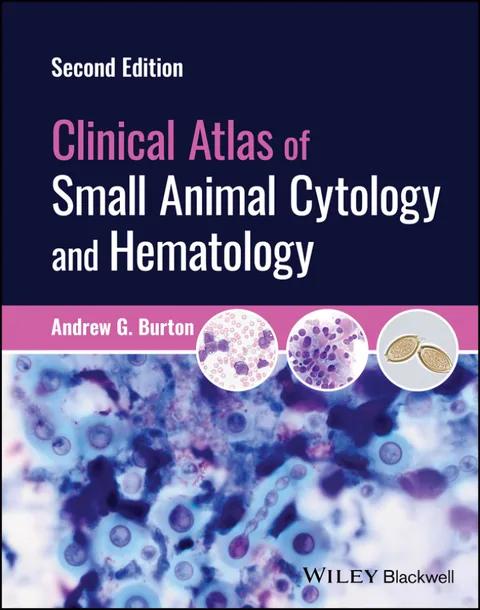
Clinical Atlas of Small Animal Cytology and Hematology
A comprehensive image-based reference to all aspects of small animal cytology and hematology, including sample collection and handling
by Andrew G. Burton
2nd Edition
Publisher: Wiley-Blackwell
Book Details
| Print ISBN | 9781119985624 |
| eText ISBN | 9781119985648 |
| Publisher | Wiley-Blackwell |
| Publishing Year | 2024 |
| Edition | 2nd Edition |
| Language | English |
| Pages | 576 |
The Clinical Atlas of Small Animal Cytology and Hematology, 2nd Edition provides a comprehensive image-based reference for diagnostic microscopic evaluation in veterinary clinical pathology. Published by Wiley-Blackwell, this reference work organizes cellular findings to assist clinicians when examining patient samples and making accurate diagnostic assessments.
Coverage spans the technical workflow of diagnostic laboratory procedures, beginning with cytology sampling, specimen preparation, staining methods, sample handling, and long-term slide storage. Organ-focused sections systematically detail microscopic features across body systems, incorporating specific coverage of the hemolymphatic system through detailed analysis of lymph nodes, spleen, thymus, and bone marrow. Additional diagnostic chapters evaluate body cavity fluids, infectious agents, integument, and musculoskeletal structures.
Microscopic interpretation is supported throughout the reference volume by over 750 high-quality images of cytology and hematology specimens depicting cellular morphology. This image-driven structure delivers focused reference support for veterinary professionals examining clinical specimens in practice or laboratory settings.
Table of Contents
Chapter 1: Cytology Sampling and Preparation
- • 1.1 Cytology
- • 1.2 Sample Collection and Preparation
- • 1.3 Sample Staining
- • 1.4 Sample Handling and Storage
Chapter 2: Cytologic Analysis of Cells
- • 2.1 Approach to Cytology Samples
- • 2.2 Sample Quality and Background
- • 2.3 Cell Types
- • 2.4 Cell Shape, Distribution, and Features
- • 2.5 Benign Versus Malignant
Chapter 3: Infectious Agents
- • 3.1 Fungi
- • 3.2 Oomycetes
- • 3.3 Algae
- • 3.4 Mesomycetozoea
- • 3.5 Protozoa
- • 3.6 Helminths
- • 3.7 Bacteria
- • 3.8 Ectoparasites
Chapter 4: Integument
- • 4.1 Cutaneous and Subcutaneous Lesions
Chapter 5: Hemolymphatic
- • 5.1 Lymph Nodes
- • 5.2 Spleen
- • 5.3 Thymus
- • 5.4 Bone Marrow
Chapter 6: Body Cavity Fluids
- • 6.1 General Classification
- • 6.2 Specific Effusions
Chapter 7: Musculoskeletal
- • 7.1 Bone
- • 7.2 Joints
- • 7.3 Muscle
Chapter 8: Hepatobiliary
- • 8.1 Liver
- • 8.2 Biliary Tract
Chapter 9: Digestive System
- • 9.1 Salivary Glands
- • 9.2 Stomach/Intestines
- • 9.3 Feces
- • 9.4 Pancreas
Chapter 10: Urinary
- • 10.1 Kidney
- • 10.2 Bladder
- • 10.3 Urine
- • 10.4 Urinary Crystals
- • 10.5 Urinary Casts
Chapter 11: Respiratory
- • 11.1 Nasal Cavity
- • 11.2 Lung
- • 11.3 Bronchoalveolar Lavage/Transtracheal Wash
Chapter 12: Endocrine
- • 12.1 Thyroid
- • 12.2 Parathyroid
- • 12.3 Chemoreceptor Tumors
- • 12.4 Adrenal Gland
- • 12.5 Pituitary Gland
Chapter 13: Reproductive
- • Male
- • 13.1 Testes
- • 13.2 Semen Analysis
- • 13.3 Prostate
- • 13.4 Penis
- • Female
- • 13.5 Ovary
- • 13.6 Mammary Glands
- • 13.7 Vaginal Cytology
Chapter 14: Neurologic
- • 14.1 Brain
- • 14.2 Cerebrospinal Fluid
- • 14.3 Spinal Cord
Chapter 15: Ocular and Special Senses
- • 15.1 Eyes: Cornea
- • 15.2 Eyes: Conjunctiva
- • 15.3 Ears
Chapter 16: Blood Smear Preparation and Evaluation
- • 16.1 The Importance of Blood Smear Evaluation
- • 16.2 Making a Blood Smear
- • 16.3 Blood Smear Staining and Handling
- • 16.4 Blood Smear Evaluation
- • 16.5 Hematology Procedures and Techniques
Chapter 17: Erythrocytes
- • 17.1 Approach to Evaluating Red Blood Cells
- • 17.2 Red Blood Cell Distribution
- • 17.3 Red Blood Cell Morphology
- • 17.4 Red Blood Cell Inclusions
- • 17.5 Red Blood Cell Neoplasia
- • 17.6 Red Blood Cell Infectious Agents
Chapter 18: Leukocytes
- • 18.1 Approach to Evaluating Leukocytes
- • 18.2 Neutrophils
- • 18.3 Neutrophil Inclusions
- • 18.4 Eosinophils
- • 18.5 Basophils
- • 18.6 Mast Cells
- • 18.7 Monocytes
- • 18.8 Lymphocytes
- • 18.9 Leukocyte Neoplasia
- • 18.10 Leukocyte Infectious Agents
Chapter 19: Platelets
- • 19.1 Approach to Evaluating Platelets
- • 19.2 Platelet Distribution
- • 19.3 Platelet Morphology
- • 19.4 Platelet Neoplasia
- • 19.5 Platelet Infectious Agents
Chapter 20: Background Features and Miscellaneous Cells
- • 20.1 Approach to Blood Smear Background Features
- • 20.2 Acellular Elements
- • 20.3 Miscellaneous Cells
- • 20.4 Infectious Agents
Customer Reviews
0.0
0 reviews
No reviews yet. Be the first to review this book!
Write a Review
Reviewed by GradeFocus Editorial Team
▶Research Sources (13)
- Clinical Atlas of Small Animal Cytology and Hematology
- Sourcebooks, LLC.
- Clinical atlas of small animal cytology and hematology
- Bound To Stay Bound Books, Inc. - Bookstore
- Title
- Clinical Atlas of Small Animal Cytology and Hematology
- Pearson eText for Discovering the Life Span, 5e - Exchange
- Civil Procedure: A Coursebook (Connected eBook with ...
- Clinical Atlas of Small Animal Cytology and Hematology, ...
- [📚New Cytology Book Alert!] Clinical Atlas of Small Animal ...
- Clinical Atlas of Small Animal Cytology and Hematology
- Atlas of diagnostic cytology in small animal - Veterinary book
- Clinical Atlas of Small Animal Cytology and Hematology
Related Books
Veterinary Necropsy Guide
Sean P. McDonough

Pathology of Laboratory Rodents and Rabbits
Stephen W. Barthold

Seizures in Dogs and Cats
Sean Sanders

Veterinary Euthanasia Techniques
Kathleen A. Cooney

Companion Animal End of Life Care for the Veterinary Technician
Alyssa Eslinger

Veterinary Oral and Maxillofacial Pathology
Brian G. Murphy