Handbook of X-ray Imaging
Physics and Technology
by Paolo Russo
1st Edition
Publisher: CRC Press
Book Details
| Print ISBN | 9781498741521 |
| eText ISBN | 9781498741552 |
| Publisher | CRC Press |
| Publishing Year | 2018 |
| Edition | 1st Edition |
| Language | English |
| Pages | 1315 |
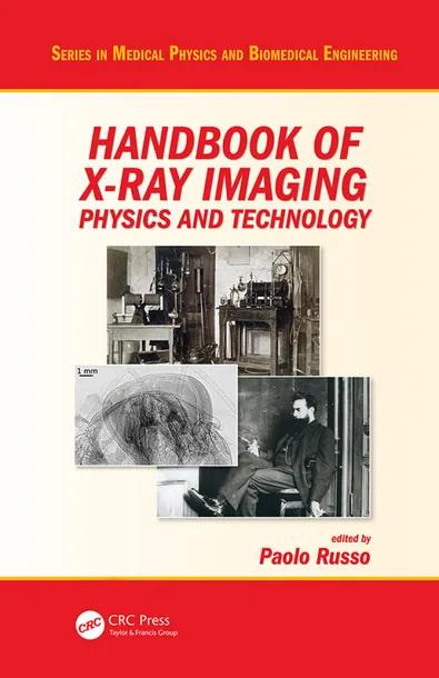
The Handbook of X-ray Imaging: Physics and Technology, 1st Edition, edited by Dr. Paolo Russo, provides an extensive reference on the physical principles and technological instrumentation of X-ray imaging. Prepared with input from an international scientific board, the handbook brings together contributions from over 130 expert contributors.
The volume addresses two- and three-dimensional imaging methods across energy spectra ranging from soft X-rays to megavoltage levels. Core topics span computed tomography, fluoroscopy, dental imaging, phase contrast imaging, and non-destructive industrial testing.
This handbook supports graduate students in medical physics and biomedical engineering, medical physics residents, radiographers, and industrial testing specialists by connecting fundamental radiation physics with clinical and technical practice.
Table of Contents
Chapter 1: Basic physics of X-ray interactions in matter
Chapter 2: Xray tube physics and technology
Chapter 3: X-ray generators
Chapter 4: Carbon Nanotube Based Field Emission X-ray Technology
Chapter 5: Technology of Miniature X-Ray Tubes
Chapter 6: Technology of pyroelectric X-ray tubes
Chapter 7: History of X-Ray Tubes
Chapter 8: Synchrotron radiation x-ray sources for radiography and tomography
Chapter 9: X-ray shutters
Chapter 10: Calculation of x-ray spectra
Chapter 11: Radiochromic film dosimetry for radiology
Chapter 12: Computed Radiography
Chapter 13: Photon Counting Detectors for X-ray imaging
Chapter 14: Image Quality
Chapter 15: Image quality in attenuation based and phase contrast based X-ray imaging
Chapter 16: Inverse Compton Scattering X-ray sources
Chapter 17: Wilhelm Conrad Roentgen - The discovery of X-rays and the creation of a new medical profession
Chapter 18: History of Radiology
Chapter 19: Digital Mammography
Chapter 20: Digital Breast Tomosynthesis
Chapter 21: Fluoroscopy: physics and technology
Chapter 22: Dental radiography
Chapter 23: Clinical mammographic and tomosynthesis units
Chapter 24: Physical image quality evaluation of x-ray detectors for digital radiography and mammography
Chapter 25: Mammography, breast tomosynthesis and risk of radiation-induced breast cancer
Chapter 26: Clinical radiographic units
Chapter 27: Clinical Fluoroscopy units
Chapter 28: Physical Basis of X-ray Breast Imaging
Chapter 29: Radiation dose in X-ray mammography and digital breast tomosynthesis
Chapter 30: Industrial Radiography
Chapter 31: Forensic Radiology
Chapter 32: X-ray Computed Tomography for diagnostic imaging – from single-slice to multi-slice
Chapter 33: Analytical reconstruction methods in X-ray CT
Chapter 34: Iterative reconstruction methods in X-ray CT
Chapter 35: X-ray Cone-Beam Computed Tomography
Chapter 36: Small Animal X-ray Computed Tomography
Chapter 37: Quality Assurance of X-ray Computer Tomography
Chapter 38: Radiation Dose in X-ray Computed Tomography
Chapter 39: Dual energy X-ray computed tomography
Chapter 40: Soft x-ray tomography: techniques and applications
Chapter 41: 4-D X-ray Computed Tomography
Chapter 42: Dental and Maxillofacial Cone Beam Computed Tomography
Chapter 43: High Speed X-ray computed tomography
Chapter 44: Kilovoltage and Megavoltage Imaging in Radiotherapy
Chapter 45: Industrial X-ray computed tomography
Chapter 46: Industrial X-ray computed tomography scanners
Chapter 47: Dimensional metrology for industrial Computed Tomography
Chapter 48: Influence of scatter in X-ray imaging and scatter correction methods for industrial applications
Chapter 49: Theory of X-ray phase-contrast imaging
Chapter 50: Non-interferometric techniques for X-ray phase-contrast biomedical imaging
Chapter 51: X-ray phase-contrast mammography
Chapter 52: X-ray phase contrast tomosynthesis imaging
Chapter 53: Crystal analyser-based X-ray Phase Contrast Imaging
Chapter 54: X-Ray scattering: analytical applications and imaging
Chapter 55: Tissue Substitute Materials for Diagnostic X-ray Imaging
Chapter 56: Phantoms for image quality and dose assessment
Chapter 57: Software phantoms for X-ray radiography and tomography
Chapter 58: Radiography and Computed Tomography for Works of Art
Chapter 59: Computer Aided Diagnosis for X-ray imaging
Chapter 60: Computer analysis of mammograms
Chapter 61: Databases for mammography
Chapter 62: Computer analysis of CT images for lung nodule detection
Chapter 63: Display Optimization & Human Factors
Chapter 64: Display for Medical Imaging and DICOM Grayscale Standard Display Function Fundamentals
Chapter 65: Quality Control of Medical Imaging Displays
Chapter 66: Radiation protection issues in X-ray radiology, fluoroscopy and computed tomography
Chapter 67: Educational aspects in radiography physics and technology
Chapter 68: Tables of X-rays mass attenuation coefficients, of K and L –energy, of K, L and M fluorescence yield, of Kα/Kβ, Lα/Lβand Lα/Lγ
Customer Reviews
0.0
0 reviews
No reviews yet. Be the first to review this book!
Write a Review
Reviewed by GradeFocus Editorial Team