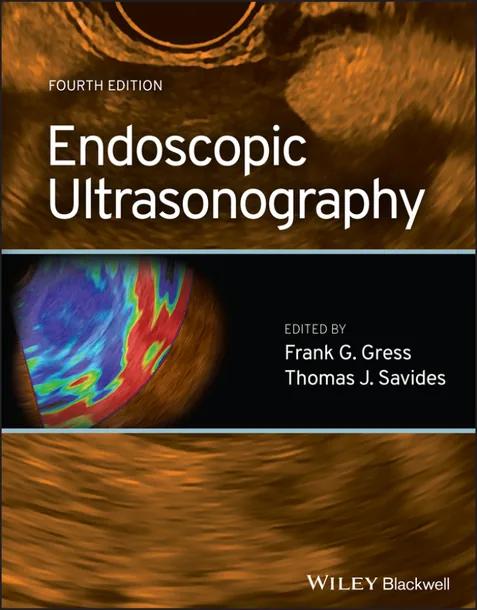
Endoscopic Ultrasonography
by Frank G. Gress, Thomas J. Savides
4th Edition
Publisher: Wiley-Blackwell
Book Details
| Print ISBN | 9781119697916 |
| eText ISBN | 9781119697930 |
| Publisher | Wiley-Blackwell |
| Publishing Year | 2024 |
| Edition | 4th Edition |
| Language | English |
| Pages | 320 |
Endoscopic Ultrasonography, 4th Edition, edited by Frank G. Gress and Thomas J. Savides, presents a comprehensive textbook focused on diagnostic, staging, and interventional endosonographic methods. Designed for gastrointestinal endoscopists and medical professionals working with gastrointestinal pathologies, the text connects baseline ultrasound principles with active procedural execution.
The content follows a clear instructional progression across technical modalities and anatomic targets. Early chapters address fundamental imaging mechanics, linear-array normal anatomy, and high-frequency ultrasound probes. The coverage then expands into specialized clinical applications, detailing mediastinal endosonography, esophageal cancer staging, stomach and duodenal evaluations, and gastrointestinal subepithelial mass assessments.
A major emphasis of the volume involves invasive tissue sampling techniques. Step-by-step guidance outlines EUS fine-needle aspiration and fine-needle biopsy protocols. In addition, dedicated sections explain cytology material preparation, cytology interpretation, and EUS elastography to support procedural accuracy in diagnostic settings.
Table of Contents
Chapter 1: A history of endoscopic ultrasonography
- • PART 1. Early history (edited by Michael Wallace)
- • Acknowledgments
- • Reference
Chapter 2: Basic principles and fundamentals of EUS imaging
- • How US images are made
- • How US waves are made
- • What happens when US waves encounter tissue
- • How images are made from reflected US waves
- • How transducer properties affect the image
- • US frequency and axial resolution
- • Transducer size and lateral resolution
- • Attenuation and tissue penetration
- • How tissue properties affect images: the GI wall
- • Frequency dependence
- • Specular and nonspecular reflectors
- • Nonspecular reflectors (scatterers)
- • Specular reflectors (interface echoes)
- • Detection of tissue movement: doppler imaging
- • New techniques in EUS imaging
- • Contrast‐enhanced EUS imaging
- • Elastography
- • Imaging artifacts
- • Reverberation artifacts
- • Tangential scanning
- • Attenuation artifacts
- • Side‐lobe artifacts
- • Doppler artifacts
- • Conclusion
- • References
Chapter 3: Learning EUS anatomy
- • General principles of EUS
- • Echo endoscopes
- • Regional anatomy
- • The esophagus and extraesophageal spaces
- • The stomach and the extragastric spaces
- • The duodenum and extraduodenal spaces
- • The rectum and the extrarectosigmoid spaces
- • Approach to understanding EUS anatomy
- • Conclusion
- • Reference
Chapter 4: EUS instruments, room setup, and assistants
- • EUS instruments and other equipment
- • Room setup
- • EUS assistants
- • Conclusion
- • References
Chapter 5: EUS procedure: consent and sedation
- • Consent
- • Infection
- • Bleeding
- • Perforation
- • FNA needle‐track seeding
- • Esophageal dilation for facilitation of EUS evaluation
- • Pancreatitis
- • Bile peritonitis
- • Specific issues related to celiac plexus neurolysis
- • Specific issues related to EUS‐guided biliary and pancreatic access
- • Sedation
- • Pre‐procedure assessment
- • Benzodiazepines
- • Opiates
- • Adjuncts to benzodiazepines and opiates
- • Propofol
- • Procedural monitoring
- • Post‐procedural monitoring
- • Conclusion
- • References
Chapter 6: The EUS report
- • Roles of the endoscopic report
- • Clinical care
- • Quality control
- • Clinical research
- • Administrative and legal issues
- • Evolution of the medical report
- • Standard terminology and structured reporting
- • Standard terminology
- • Structured reporting
- • Speed
- • Completeness
- • Free text and conventional reports
- • Databases
- • Hierarchical
- • Network
- • Relational
- • Commercial software for EUS reporting
- • The EUS report
- • Non‐EUS information
- • General EUS information
- • EUS interventions (diagnostic and therapeutic)
- • Complications
- • Procedure summary
- • Quality of EUS reports
- • Disease‐specific information
- • Luminal cancer (esophageal, gastric, rectal)
- • Subepithelial lesions
- • Solid pancreatic tumor
- • Pancreatic cystic lesions
- • Pancreatitis
- • Conclusion
- • References
Chapter 7: Endosonography of the mediastinum
- • Case
- • Introduction
- • What are the techniques for performing EUS/EUS‐FNA in the mediastinum?
- • When is EUS/EUS‐FNA of the mediastinum indicated?
- • Diagnosis
- • Malignant diseases
- • Interventional endoscopy
- • Summary
- • Complications
- • Conclusion
- • References
Chapter 8: Linear‐array EUS: normal anatomy
- • Performing the examination
- • The linear esophagus
- • The linear stomach
- • The linear duodenum
- • The linear rectum
- • Male
- • Female
- • Conclusion
- • References
Chapter 9: High‐frequency ultrasound probes
- • High‐frequency ultrasonography
- • Technical features
- • Anatomical correlation
- • Applications of high‐frequency ultrasonography
- • Esophagus
- • Esophageal cancer
- • Barrett esophagus
- • Other indications
- • Stomach
- • Early gastric cancer
- • Small bowel and colon
- • Colorectal cancer
- • Other applications
- • Intraductal ultrasonography
- • Applications of IDUS
- • Choledocholithiasis
- • Bile duct strictures
- • Cholangiocarcinoma
- • Ampullary tumors
- • Pancreatic adenocarcinoma and pancreatic strictures
- • Mucin‐producing tumors
- • Complications
- • The future
- • Conclusion
- • References
Chapter 10: EUS elastography
- • Introduction
- • Technical aspects and methodology of elastography
- • Strain elastography
- • Qualitative strain elastography
- • Quantitative strain elastography
- • Shear wave elastography
- • Clinical applications of EUS elastography
- • Pancreatic diseases
- • Differential diagnosis of solid pancreatic lesions
- • Chronic pancreatitis
- • Lymph nodes
- • Gastrointestinal lesions
- • Transrectal EUS elastography
- • Other indications
- • Conclusion
- • References
Chapter 11: Fundamentals of EUS FNA
- • Pre‐procedural fundamentals
- • Indications and contraindications of EUS FNA
- • Establishing procedural goals
- • Informed consent
- • Prophylactic antibiotics
- • Equipment and staffing
- • Training
- • Intraprocedural fundamentals
- • Algorithm to obtain EUS FNA
- • Accessing the target lesion
- • EUS FNA technique
- • Lesion characteristics
- • Needle size
- • Use of suction during EUS FNA
- • Use of a stylet during EUS FNA
- • Helpful tips for obtaining adequate EUS FNA specimens
- • Role of onsite cytopathology
- • Post‐procedural fundamentals
- • Monitoring of the patient
- • Safety of EUS FNA
- • Fine needle biopsy (FNB)
- • Conclusion
- • References
Chapter 12: EUS FNA cytology: material preparation and interpretation
- • Introduction
- • Technical preparation and quality of EUS biopsy material
- • Technical quality of EUS biopsy material
- • Personnel
- • Cell block
- • Needle size
- • Needle preparation
- • Suction
- • Slide preparation and staining
- • Liquid‐based preparations
- • Usefulness of pancreatic cyst fluid analysis
- • Molecular analysis
- • Quality of the interpretation
- • Integration of pathologic and clinical information
- • Rapid onsite cytologic evaluation
- • Role of the laboratory in EUS
- • References
Chapter 13: EUS: applications in the mediastinum
- • Mediastinal cysts
- • Mediastinal cyst aspiration or not mediastinal cyst aspiration, that is the question
- • Lung cancer
- • Rationale for EUS
- • Before you start
- • Cross‐sectional and functional imaging: how does EUS stack up?
- • Medical mediastinoscopy
- • Endobronchial ultrasonography
- • EUS in early NSCLC
- • Failed bronchoscopy and EUS rescue
- • EUS and MS
- • Getting the examination done
- • Radial EUS
- • Linear EUS
- • Which lymph nodes for FNA?
- • FNA: how and how much?
- • Special topics
- • Primary lung lesions
- • T4 disease
- • EUS for metastatic disease
- • EUS after neoadjuvant therapy
- • Cost
- • Training
- • Conclusion
- • References
Chapter 14: EUS for esophageal cancer
- • Introduction
- • Staging Guidelines for Esophageal Cancer
- • Role of EUS in the Staging of Esophageal Cancer
- • Staging of Esophageal Cancer Using EUS
- • Impact of EUS Staging on Management
- • Limitations of EUS
- • References
Chapter 15: EUS of the stomach and duodenum
- • Benign disorders
- • Menetrier's disease and other hypertrophic gastropathies
- • Gastric varices
- • Nonhealing gastric ulcer
- • Perigastric/Periduodenal lesions
- • Therapeutic EUS of the stomach and duodenum
- • Malignant disorders
- • Gastric cancer
- • Intestinal‐type gastric cancer
- • Early gastric cancer (intestinal type)
- • Diffuse‐type gastric cancer
- • Gastric lymphoma
- • Diffuse large B‐cell‐type non‐Hodgkin lymphoma
- • MALT lymphoma (extranodal marginal‐zone B‐cell lymphoma)
- • EUS staging of gastric lymphoma
- • Detection of ascites
- • Benign lesions of the duodenum, ampullary adenomas, and ampullary carcinoma
- • Duodenal adenomas
- • Ampullary lesions
- • Conclusion
- • References
Chapter 16: EUS for gastrointestinal subepithelial masses
- • Endoscopic findings
- • EUS imaging techniques
- • Esophagus
- • Stomach
- • Duodenum
- • Rectum and colon
- • Lesions located in the mucosal layer
- • Mucosal polyps
- • Mucosal cysts
- • GI stromal tumors or leiomyomas
- • Lesions located in the submucosa
- • Lipomas
- • Carcinoid tumors
- • Granular cell tumors
- • Duplication cysts
- • Heterotopic pancreas (pancreatic rest or ectopic pancreas)
- • Varices
- • Lesions located in the muscularis propria
- • Stromal cell tumors (GISTs and leiomyomas)
- • Duodenal gangliocytic paraganglioma
- • Duodenal adenomyomatosis
- • Extrinsic compression lesions
- • Comparison of imaging studies for subepithelial masses
- • Utility of EUS in the management of subepithelial masses
- • Interobserver agreement of the EUS in evaluating submucosal masses
- • Outcome studies
- • Endoscopic tissue sampling
- • Which lesions need sampling?
- • Biopsy forceps
- • Paradigm shift from EUS‐guided FNA to FNB
- • Removal of overlying mucosa, followed by a deep biopsy
- • EMR, ESD, STER, and EFTR
- • Endoscopic and laparoscopic resection of submucosal masses
- • Conclusion
- • References
Chapter 17: EUS and its emerging modalities for the diagnosis and staging of pancreatic tumors
- • Endoscopic ultrasonography
- • EUS‐fine needle aspiration (EUS‐FNA)
- • Contrast‐enhanced EUS
- • Elastography
- • Staging pancreatic cancer with different techniques
- • Conclusion
- • References
Chapter 18: EUS for pancreatic cysts
- • Pancreatic cyst classification
- • EUS morphology
- • EUS‐guided FNA and cyst fluid analysis
- • Biochemical analysis
- • Molecular analysis
- • Histologic analysis
- • Characteristics of pancreatic cystic lesions
- • Pancreatic cystic neoplasms
- • Mucinous cystic neoplasms
- • Intraductal papillary mucinous neoplasms
- • Serous neoplasms
- • Solid pseudopapillary neoplasms
- • Nonneoplastic pancreatic cysts
- • Inflammatory pancreatic fluid collections
- • Solid tumors presenting as pancreatic cysts
- • EUS‐FNA technique
- • Evolving approaches
- • Conclusion
- • References
Chapter 19: The role of diagnostic EUS in inflammatory diseases of the pancreas
- • Acute pancreatitis
- • Recurrent acute pancreatitis
- • Chronic pancreatitis
- • EUS features of CP and reproducibility
- • EUS inter‐ and intra‐observer variability in CP
- • Rosemont criteria
- • Confounders associated with EUS as a diagnostic tool for CP
- • Factors Influencing EUS Findings in the Pancreas
- • Abnormal EUS features in patients with clinical symptoms but normal imaging and/or pancreatography
- • Abnormal EUS in patients without clinical symptoms
- • Comparison of EUS findings for CP with other diagnostic modalities
- • Comparison to histology
- • Comparison to ERCP and secretin‐stimulated duodenal aspiration
- • Comparison to CT and TUS
- • Comparison to MRCP
- • EUS‐guided tissue sampling for diagnosis of CP
- • Differentiating CP and pancreatic cancer
- • Developing EUS technologies for the diagnosis of CP
- • Elastography
- • Contrast‐enhanced EUS
- • Digital image analysis
- • Conclusion
- • References
Chapter 20: Autoimmune pancreatitis
- • Classification of AIP
- • Clinical presentation of AIP
- • Diagnosis of AIP
- • Diagnosis of type 1 AIP
- • Diagnosis of type 2 AIP
- • Distinguishing AIP from pancreatic cancer
- • Role of other tests in AIP
- • CT and MRI
- • Endoscopic retrograde cholangiopancreatography
- • Ampullary biopsy
- • EUS imaging features of AIP
- • EUS imaging of the pancreas
- • EUS imaging of other organs
- • Image enhancement techniques in EUS
- • EUS‐guided tissue acquisition
- • EUS‐FNA
- • EUS‐FNB
- • Treatment and outcomes of AIP
- • Conclusion
- • References
Chapter 21: EUS for biliary diseases
- • Common bile duct stones
- • Acute biliary pancreatitis
- • Indeterminate biliary strictures
- • Gallbladder polyps and cancer
- • EUS‐guided biliary drainage
- • EUS‐guided gallbladder drainage
- • Conclusion
- • Acknowledgment
- • References
Chapter 22: EUS in liver disease
- • EUS imaging technique to evaluate the liver
- • Liver parenchymal abnormalities
- • Liver cirrhosis and portal hypertension assessments
- • Hepatic steatosis
- • EUS‐guided liver biopsy
- • EUS‐guided portal pressure gradient measurements
- • Emerging imaging techniques: artificial intelligence and virtual biopsy
- • Malignant lesions in the liver
- • Hepatocellular carcinoma and other primary liver tumors
- • Liver metastases
- • Benign lesions in the liver
- • Hepatic adenomas
- • Focal nodular hyperplasia
- • Hemangiomas
- • Cystic liver lesions
- • Intrahepatic biliary disorders
- • Intrahepatic cholangiocarcinoma
- • Primary sclerosing cholangitis
- • Biliary adenomas
- • Conclusions
- • References
Chapter 23: Colorectal EUS
- • Introduction
- • Instruments for colorectal endosonography
- • Rigid probes
- • Echoendoscopes
- • Examination technique
- • Colorectal cancer staging by EUS
- • Tumor (T) stage
- • Lymph node (N) stage
- • Distant metastastis (M) stage
- • Accuracy of T‐staging
- • Accuracy of N‐staging
- • Fine‐needle aspiration (FNA)
- • Interobserver variability in rectal cancer staging by EUS
- • EUS compared to CT and MRI
- • Three‐dimensional EUS for rectal cancer staging
- • Contrast‐enhanced EUS for rectal cancer staging
- • Clinical impact of EUS staging in rectal cancer
- • EUS for local recurrence of colorectal carcinoma
- • Restaging after chemotherapy and radiation
- • Linitis plastica of the rectum
- • Anal cancer
- • Anal sphincter defects
- • Subepithelial lesions and compression of the colorectal wall
- • Rectosigmoid and pelvic endometriosis
- • I‐anorectal abscess and fistula
- • Endoscopic ultrasound in inflammatory bowel disease beyond imaging for perianal fistulas
- • EUS‐guided drainage of perirectal abscesses
- • Prostate cancer and rectal EUS
- • Other pelvic malignancies
- • Conclusion
- • References
Chapter 24: Therapeutic EUS for cancer treatment
- • EUS‐guided delivery of antitumor agents
- • EUS‐guided tumor ablation
- • EUS RFA and PDT
- • EUS‐guided laser ablation
- • EUS‐guided placement of fiducial markers and brachytherapy
- • EUS‐guided celiac neurolysis
- • Technique
- • Results
- • Conclusion
- • References
Chapter 25: EUS‐guided biliary access
- • Introduction
- • Equipment
- • Echoendoscope
- • EUS needles
- • Other instruments
- • Indications
- • Technique
- • Stent selection
- • Outcome
- • Procedure failure and optimizing the technique
- • Conclusion
- • References
Chapter 26: Pancreatic fluid collections
- • Abbreviations
- • Introduction
- • Classification of pancreatic fluid collections
- • Treatment of pancreatic fluid collections
- • Surgical drainage
- • Percutaneous drainage
- • Endoscopic drainage
- • Conventional versus EUS‐guided endoscopic drainage
- • Choice of stent in endoscopic drainage
- • Comparison studies between the different drainage modalities
- • Walled‐off pancreatic necrosis (WOPN)
- • Direct endoscopic necrosectomy (DEN)
- • Surgical necrosectomy
- • Comparison studies between the techniques
- • Enteral feeding
- • Pancreatic ductal evaluation
- • Tailored multidisciplinary approach based on the creation of an algorithm
- • Conclusion
- • References
Chapter 27: EUS‐guided enteric anastomoses
- • Introduction
- • EUS‐guided gastroenterostomy for gastric outlet obstruction
- • EUS‐gastroenterostomy: technical aspects and clinical outcomes
- • Safety of EUS‐gastroenterostomy for gastric outlet obstruction
Customer Reviews
0.0
0 reviews
No reviews yet. Be the first to review this book!
Write a Review
Reviewed by GradeFocus Editorial Team
▶Research Sources (14)
- [PDF] Endoscopic Ultrasonography by Frank G. Gress, 4th edition - Perlego
- books by Frank G. Gress author | BookScouter.com
- Store - Clearwater Press
- Endoscopic Ultrasonography | Rent | 9781119697916 - eCampus.com
- Endoscopic ultrasonography
- books by Thomas J. Savides author | BookScouter.com
- Welcome to the AMLE Store
- Endoscopic Ultrasonography 2016 Third edition;3rd Edition
- Endoscopic Ultrasonography Edition:4th ISBN ...
- Endoscopic Ultrasonography [4 ed.] 1119697913, ...
- Endoscopic ultrasound: what is it and when should it be used?
- Endoscopic Ultrasonography : Frank G. Gress (editor)
- Endoscopic Ultrasonography, 4th Edition - 9781119697916
- Endoscopic Ultrasonography, 4th Edition by Frank G. Gress
Related Books

Translational Gastroenterology
Adam E.M. Eltorai
Handbook of Gastrointestinal Motility and Disorders of Gut-Brain Interactions
Satish S.C. Rao

Sleisenger and Fordtran's Gastrointestinal and Liver Disease
Mark Feldman
Sleisenger and Fordtran's Gastrointestinal and Liver Disease Review and Assessment
Emad Qayed
Sitaraman and Friedman's Essentials of Gastroenterology
Shanthi Srinivasan
Yamada's Textbook of Gastroenterology
Timothy C. Wang